
Brain Imaging Study Reveals Unexpected Glymphatic Pattern in Adolescent Psychosis
Brain Imaging Study Reveals Unexpected Glymphatic Pattern in Adolescent Psychosis https://pediatricsnationwide.org/wp-content/uploads/2024/04/AdobeStock_141694591-1024x683.jpg 1024 683 Katie Brind'Amour, PhD, MS, CHES https://pediatricsnationwide.org/wp-content/uploads/2021/03/Katie-B-portrait.gif
Higher glymphatic imaging markers in adolescents contrast with patterns reported in adult studies, highlighting the importance of developmental stage for biomarker interpretation.
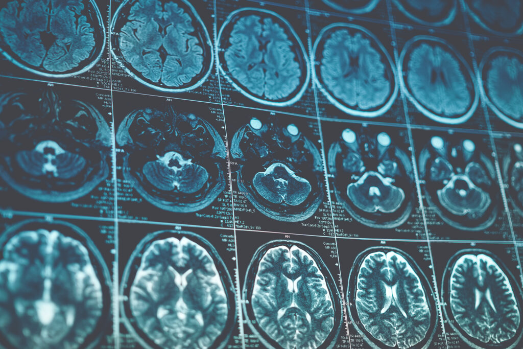
A new neuroimaging study from investigators at Nationwide Children’s Hospital suggests that adolescents with first-episode of psychosis (FEP) may have increased activity in the brain’s glymphatic (waste clearance) system as measured by an MRI marker, in contrast to multiple studies of adults with schizophrenia and chronic psychosis.
The study compared 21 adolescent patients with FEP with 42 age- and sex-matched community controls. Using diffusion MRI, they measured the ALPS (Analysis Along the Perivascular Space) index, an indirect MRI marker related to glymphatic function. They also found significantly lower cortical thickness in the psychosis group, consistent with previous research showing accelerated cortical thinning in youth with psychotic disorders.
“Cortical thinning is a normal developmental process, but we know the trajectory is different in kids who develop psychosis,” says Eric E. Nelson, PhD, co-author of the study, principal investigator and director of neuroimaging research in the Center for Biobehavioral Health at Nationwide Children’s. “One longstanding theory is that synaptic pruning becomes overly aggressive during adolescence, removing connections that are important for healthy brain function. We don’t think the glymphatic system is doing the pruning itself, but it may be a marker of how much remodeling is happening and how much material needs to be cleared from the brain.”
The glymphatic system functions as the brain’s waste-clearance network, helping remove proteins and metabolic byproducts through fluid pathways surrounding blood vessels. While ALPS is not a direct measure of glymphatic function, it is increasingly being explored as a noninvasive imaging biomarker.
Because pre-pubertal psychosis is extremely rare, adolescence may represent a critical neurodevelopmental window for understanding how psychosis emerges and how biomarkers could aid earlier detection and intervention.
“Our findings show that developmental stage may fundamentally affect how we interpret biological markers, and that the adolescent brain is not simply a smaller version of the adult brain,” says Musa Yilanli, MD, child and adolescent psychiatrist at Nationwide Children’s and senior author of the study. “These findings may reflect developmental stage, illness stage, medication exposure or other differences between study populations — we need longitudinal studies to understand why the pattern may differ in adolescents. It’s a valuable window into what the brain may look like closer to the onset of disease.”
The study also explored potential connections between glymphatic activity and sleep-related symptoms and psychosis symptom severity, but did not find significant associations. The team cautions against the immediate endorsement of ALPS as a clinical biomarker, but is continuing to employ it in ongoing studies in adolescents experiencing suicidal thoughts and behaviors and youth with other psychiatric conditions.
“This study gives us an important signal, but it is just a starting point,” says Emily Alder, PhD, lead author of the study and program manager for the Brain Development and Social Cognition Lab in the Center for Biobehavioral Health. “ALPS is still an indirect measure of glymphatic function, and we need larger, longitudinal studies to understand what these changes mean over time. But it’s an encouraging reason to keep evaluating biomarkers that could help us better understand psychosis.”
The team also collects longitudinal blood samples and a multifaceted survey battery of cognitive and psychiatric functioning, development and family data to better understand the causes of psychosis and potential individualized treatment approaches.
“Right now, we have a photograph,” says Dr. Yilanli. “What we really need is a movie. The next step is trying to understand how these biological measures change during development, treatment and recovery, so we can find the best ways to intervene.”
Reference:
Alder E, Among K, Olexa CA, Hoskinson KR, Mattson WI, Nelson EE, Yilanli M. Increased Activity of the Glymphatic System in First-Episode Psychosis: Relationship With Cortical Thinning. JAACAP Open. 2026 Apr 23;4(4):691-700. 10.1016/j.jaacop.2026.04.004
Image credit: Adobe Stock
About the author

Katherine (Katie) Brind’Amour is a freelance medical and health science writer based in Pennsylvania. She has written about nearly every therapeutic area for patients, doctors and the general public. Dr. Brind’Amour specializes in health literacy and patient education. She completed her BS and MS degrees in Biology at Arizona State University and her PhD in Health Services Management and Policy at The Ohio State University. She is a Certified Health Education Specialist and is interested in health promotion via health programs and the communication of medical information.
- Katie Brind'Amour, PhD, MS, CHEShttps://pediatricsnationwide.org/author/katie-brindamour-phd-ms-ches/

- Katie Brind'Amour, PhD, MS, CHEShttps://pediatricsnationwide.org/author/katie-brindamour-phd-ms-ches/
- Katie Brind'Amour, PhD, MS, CHEShttps://pediatricsnationwide.org/author/katie-brindamour-phd-ms-ches/
- Katie Brind'Amour, PhD, MS, CHEShttps://pediatricsnationwide.org/author/katie-brindamour-phd-ms-ches/