
MRI Study Points to Glymphatic System Changes in Pediatric Demyelinating Disorders
MRI Study Points to Glymphatic System Changes in Pediatric Demyelinating Disorders https://pediatricsnationwide.org/wp-content/uploads/2026/10/AdobeStock_1104874692-MRI-crop-1024x445.jpg 1024 445 Pam Georgiana https://pediatricsnationwide.org/wp-content/uploads/2023/07/May-2023.jpg
Findings could help researchers understand how brain changes evolve over the course of disease.
Relatively little is known about the brain’s glymphatic system. Researchers know that it helps remove waste products from the brain, but they are still learning how this recently discovered system may be affected by neurological disease. A new pilot study from Nationwide Children’s Hospital, published in the Journal of the Neurological Sciences, suggests glymphatic function may be altered in children with acquired demyelinating disorders.
Pediatric acquired demyelinating syndromes are a group of immune-mediated disorders that damage myelin. This protective covering surrounds nerve fibers in the central nervous system. The disorders include multiple sclerosis and myelin oligodendrocyte glycoprotein antibody-associated disease (MOGAD).
Previous studies have found evidence of glymphatic system changes in certain neurological conditions, including multiple sclerosis. Research into pediatric demyelinating disorders, however, remains limited.

Florencia Ontiveros
“So far, studies have researched the different diagnoses separately,” says Florencia Ontiveros, clinical research coordinator in the Center for Biobehavioral Health in the Abigail Wexner Research Institute at Nationwide Children’s. “It was one study on patients with multiple sclerosis, and then another paper with a strictly MOGAD group. We wanted to look at all of the pediatric diagnoses together to see if we would find the same pattern.”
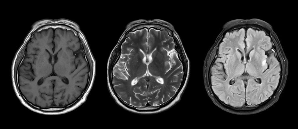
The researchers assessed glymphatic function using diffusion MRI data to measure the movement of water molecules along the spaces surrounding blood vessels in a specific region of the brain. From these measurements, they calculated the diffusion tensor image analysis along the perivascular space (DTI-ALPS) index. A lower index suggests less fluid movement, which may indicate reduced glymphatic function. DTI-ALPS provides a less invasive but indirect measure of glymphatic function in one area of the brain rather than a direct assessment of the entire system.

Eric Nelson, PhD
“The standard MRI usually just looks for lesions,” Eric E. Nelson, PhD, principal investigator in the Center for Biobehavioral Health at Nationwide Children’s and professor of pediatrics at The Ohio State University College of Medicine, says. “The DTI-ALPS could potentially become an additional measure for understanding how pediatric demyelinating disorder affects the brain.”
Compared with healthy controls, children with pediatric demyelinating disorders had significantly lower ALPS indices on both sides of the brain. Researchers did not find significant associations between ALPS indices and neurocognitive screening, lesion measures or white matter measures.
“The lower ALPS indices in children are particularly interesting because it suggests these differences can be detected relatively early in the course of demyelinating disease,” Dr. Nelson says.
The researchers also examined follow-up imaging from eight participants approximately six months after a relapse. Seven had higher ALPS indices at follow-up, although the group-level change was not statistically significant.
“Longitudinal research like this has not yet been done. I think we are one of the very first to do it,” Ontiveros says. “We hope that this observation will trigger more studies to take a look at symptomatic episodes, non-symptomatic episodes and how the ALPS fluctuates with each over time.”
Because the study was small, researchers could not determine whether glymphatic changes differ by diagnosis. DTI-ALPS is also an indirect measure of glymphatic function and should not be considered a diagnostic tool. Future studies could examine how the index changes across diagnoses and disease stages.

Kelsey E. Poisson, MD
Kelsey E. Poisson, MD, co-director of the Neuroimmunology Program and attending child neurologist and neuroimmunologist at Nationwide Children’s and assistant professor of clinical pediatrics and neurology at The Ohio State University College of Medicine, says that following children over time may be particularly informative.
“We know that children with ADS tend to recover better from relapses than do adults, and we also know that ALPS index is dynamic over the lifespan based on age,” Dr. Poisson says. “Future work evaluating serial assessments of the glymphatic system in children and adolescents may help us better understand their differing clinical trajectories from their adult counterparts and if the glymphatic system may play a role.”
Reference:
Ontiveros F, Mattson WI, Wilson CS, Poisson KE, Hoskinson KR, McAllister AS, Webb AI, Douma A, Nelson EE. Evidence for compromised function of the glymphatic system in pediatric acquired demyelinating disorders. Journal of the Neurological Sciences. 2026;485:125871. doi:10.1016/j.jns.2026.125871
Image credits: Nationwide Children’s
About the author

Pam Georgiana is a brand marketing professional and writer located in Bexley, Ohio. She believes that words bind us together as humans and that the best stories remind us of our humanity. She specialized in telling engaging stories for healthcare, B2B services, and nonprofits using classic storytelling techniques. Pam has earned an MBA in Marketing from Capital University in Columbus, Ohio.
- Pam Georgianahttps://pediatricsnationwide.org/author/pam-georgiana/

- Pam Georgianahttps://pediatricsnationwide.org/author/pam-georgiana/

- Pam Georgianahttps://pediatricsnationwide.org/author/pam-georgiana/

- Pam Georgianahttps://pediatricsnationwide.org/author/pam-georgiana/