Transforming the Approach to Cancer Epigenomic Studies
Transforming the Approach to Cancer Epigenomic Studies https://pediatricsnationwide.org/wp-content/uploads/2023/03/zebrafish-for-web-1024x764.jpg 1024 764 Abbie Miller https://pediatricsnationwide.org/wp-content/uploads/2023/05/051023BT016-Abbie-Crop.jpg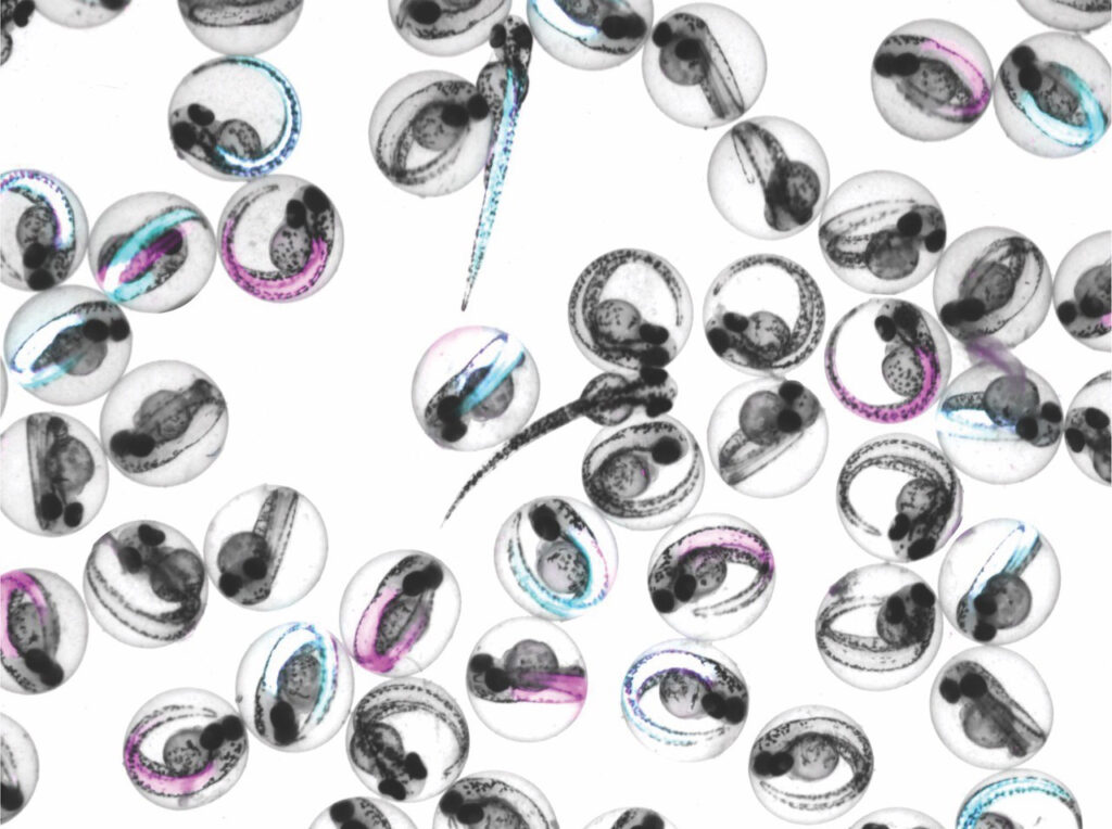
Two new publications from the Center for Childhood Cancer Research at Nationwide Children’s offer a new platform technology and proof of concept that illuminates the role of a known gene fusion driving rhabdomyosarcoma.
Synergy is an important part of scientific endeavors, and people, teams and organizations who can harness the energy of ideas and passion for the work can leverage it to move their fields forward.
At Nationwide Children’s Center for Childhood Cancer Research, such synergy has driven a collaborative project between two labs, resulting in two significant publications: one in Cell Reports and a platform technology in Cell Reports Methods for a transformative understanding of epigenetics.
Genevieve Kendall, PhD, and Benjamin Stanton, PhD, started their labs in the Center for Childhood Cancer Research in 2019. Dr. Kendall focuses her research on using zebrafish and other basic and translational models to understand pediatric sarcomas — devastating and aggressive solid tumors with limited treatment options. Dr. Stanton studies cancer epigenetics and genomics and is an expert in developing new ways to measure and understand epigenetic data.
“We started working together and the synergy was obvious: we had complementary expertise, and Dr. Stanton and I, and our groups, worked well together,” says Dr. Kendall. “In 2020-21, we got a grant from the center to really start this collaborative work in earnest. It’s been exciting to watch it grow from there.”
Now supported by National Institutes of Health and foundation funding, the teams have published two important papers as a result of their collaboration: one focused on a new methodology and one using the methodology in an embryonic zebrafish model of fusion-positive rhabdomyosarcoma (RMS).

The Kendall and Stanton Labs: seated (left to right), Andrew Vontell, Alexi Tallan, Delia Calderon, Hayden Statmore; standing (left to right), Zhiyu Song, Katie Silvius, Jack Kucinski, Cenny Taslim, PhD, Lisa Hall, DO, Rachel Hoffman, PhD, Benjamin Stanton, PhD, Genevieve Kendall, PhD, Chinmay Sankhe, PhD, Chamithi Karunanayake, PhD, Maddie Kouche.
Applying the Methodology to Understand Fusion-Positive RMS
The gene fusion PAX3::FOXO1 has been a known driver of fusion-positive RMS for more than 30 years, but to date, there are no targeted therapies. Could understanding the epigenetic events associated with the functions of the fusion oncoprotein reveal new opportunities for precision medicine? Dr. Kendall and Dr. Stanton think that quantitatively defining and understanding where the major fusion oncoproteins “lived” in the genome was the key to eventually targeting their function.
To find out, the Kendall Lab partnered with the Stanton Lab to use a new modular approach to quantitative protein-genome binding, PerCell ChIP-seq spike-in, to precisely identify binding sites and changes to the two-dimensional chromatin landscape in an embryonic zebrafish model.
“We used our newly developed PAX3::FOXO1 embryonic zebrafish model and the PerCell ChIP-seq approach to study the earliest stages of activity for one of the major fusions that drive fusion-positive rhabdomyosarcoma — PAX3::FOXO1,” Dr. Kendall explains. “This work provides important insight into in vivo mechanisms and the immediate chromatin regulatory functions of PAX3::FOXO1.”
The study found that PAX::FOXO1 reorchestrates the chromatin landscape to promote distinct neuronal types and states (neuronal transcriptional signatures) — including a neural cluster observed in fusion-positive RMS, identifying a potential target for additional research. This neural cluster is found in primary patient samples but is lost once cells are cultured in a dish, highlighting vertebrate zebrafish as an important tool to recapitulate complex in vivo biology.
“Importantly, what we’ve done here is broadly implementable and can be used to define the oncogenic and pioneering activities of known fusions with the addition of comparative and quantitative analysis,” says Dr. Kendall. “This is an important step for taking what we learn in zebrafish, mice and humans and translating it into impactful changes to care and outcomes for children with cancer.”
In addition to helping drive disease-specific discovery, the work also has implications beyond rare childhood cancers.
“The basic science work here is really impactful for pediatric cancer,” says Dr. Stanton. “But by learning about a process at the basic science level, we’ve really learned something about how nature works — and that applies broadly.”
Transforming the Approach
ChIP-seq (Chromatin ImmunoPrecipitation followed by Sequencing) is the foundational methodology for epigenetics. It maps the two-dimensional structure of chromatin inside the cell, allowing scientists to understand the unique topography of the chromatin in each sample. However, quantitative comparisons in chromatin topography between datasets remain an issue.
“Imagine the data sets are mapping the topography of mountain ranges instead of chromatin,” says Dr. Stanton. “The tool can tell us the highest peak in each sample, the lowest, and so on, but it can’t be used to compare the highest peak in the Rocky Mountains to the highest peak in the Appalachian Mountains. That quantitative cross-sample analysis holds huge opportunity for understanding epigenetic drivers of disease, and that’s what our work aimed to address.”
A spike-in approach adds DNA with known characteristics to the sample for baseline comparisons. However, the current approach to spike-ins is not designed for multicellular organisms and creates technical hurdles. This significantly limits their usability. By building on ChIP-seq and enabling a modular, intuitive, integration of wet lab and dry lab approaches, the concept of spike-ins, Drs. Stanton and Kendall and their team developed an approach that enables direct quantitative comparisons, even for complex animal systems.
PerCell ChIP-seq takes advantage of “orthologous” genomes (genes in different species that evolved from a common ancestral gene) that can be resolved through next-generation sequencing, even when indexed the same way, within the same experimental sample. The spike-in used in this approach supports efficient, cost-effective, non-commercial and reproducible strategies for epigenomic sequencing.
“This creates an opportunity for more collaboration, more analysis and a more rigorous understanding of chromatin landscapes in cancer and other disease states” says Dr. Stanton. It is so exciting that transforming technologies can illuminate epigenetic mechanisms of disease pathogenesis. Understanding the early-immediate chromatin targets of cancer-causing transcription factors will lead to new opportunities for precision therapy.”
“This collaboration has been transforming,” adds Dr. Stanton, “and we are so proud of our amazing graduate students who pioneered these studies.”
Acknowledgements: These papers were led by graduate students Alexi Tallan and Jack Kucinski as part of their dissertations. Co-authors at Nationwide Children’s for the Cell Reports and/or Cell Reports Methods paper include Ben Sunkel, Cenny Taslim, Stephanie LaHaye, Meng Wang, Andrew Vontell, Katherine Silvius, and Matthew Cannon. External co-authors for the Cell Reports Methods paper include Jun Qi and Qi Liu at Dana-Farber Cancer Institute.
References:
- Tallan A, Kucinski J, Sunkel B, Taslim C, LaHaye S, Liu Q, Qi J, Wang M, Kendall GC, Stanton BZ. Highly quantitative measurement of differential protein-genome binding with PerCell chromatin sequencing. Cell Rep Methods. 2025 Jun 16;5(6):101052.
- Kucinski J. Tallan A, Taslim C, Vontell AM, Silvius KM, Wang M, Cannon MV, Stanton BZ, Kendall GC. Rhabdomyosarcoma fusion oncoprotein initially pioneers neural signature in vivo. Cell Reports. 22 July 2025;44(7):115923. [Epub ahead of print]
Image credits: Nationwide Children’s
About the author

Abbie (Roth) Miller, MS, MWC, is a passionate communicator of science. As the manager of medical and science content at Nationwide Children’s Hospital, she shares stories about innovative research and discovery with audiences ranging from parents to preeminent researchers and leaders. She is a Medical Writer Certified®, credentialed by the American Medical Writers Association, and received her masters of science in Health Communication from Boston University.
- Abbie Millerhttps://pediatricsnationwide.org/author/abbie-miller/

- Abbie Millerhttps://pediatricsnationwide.org/author/abbie-miller/
- Abbie Millerhttps://pediatricsnationwide.org/author/abbie-miller/
- Abbie Millerhttps://pediatricsnationwide.org/author/abbie-miller/