Mapping the 3D Structure of Rhabdomyosarcoma Chromatin
Mapping the 3D Structure of Rhabdomyosarcoma Chromatin https://pediatricsnationwide.org/wp-content/uploads/2020/04/Chromatin-1140x640-1-1024x575.jpg 1024 575 Abbie Miller https://pediatricsnationwide.org/wp-content/uploads/2023/05/051023BT016-Abbie-Crop.jpg
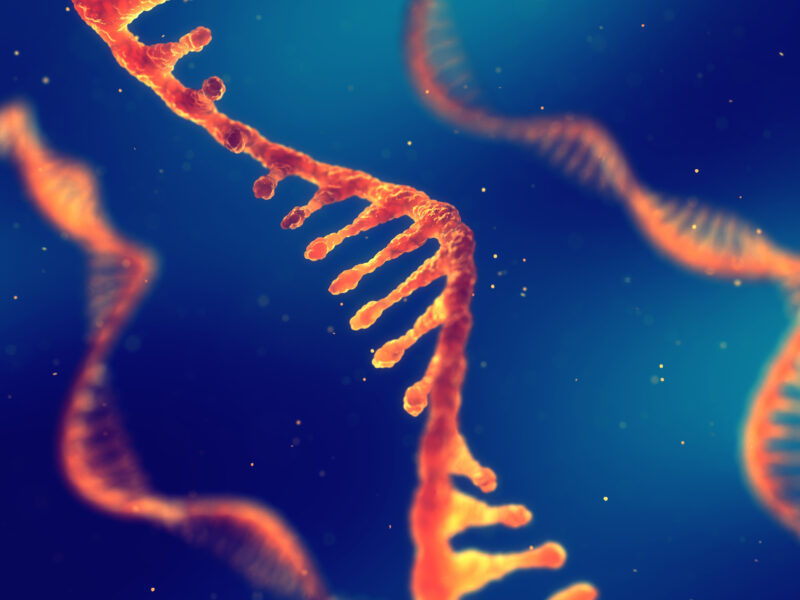
Researchers published the first comprehensive, 3D analysis of the complete rhabdomyosarcoma genome.
In a paper published in Nucleic Acids Research Cancer (NAR Cancer), researchers from the Center for Childhood Cancer Research at Nationwide Children’s and their collaborators report a comprehensive 3D chromatin structural analysis and characterization of rhabdomyosarcoma (RMS). RMS is a pediatric soft-tissue tumor that predominantly affects children between 0 and 4 years, but it can also affect adolescents. Survival rates vary dramatically from 10% to 90% depending on the stage and subtype.
“RMS represents an unmet medical need in pediatric cancer,” says Benjamin Stanton, PhD, principal investigator in the Center for Childhood Cancer Research and senior author of the publication. “Few mutations have been identified in this tumor, and even then, precision medicine remains a challenge. By understanding the epigenetics – the factors affecting gene expression outside of the actual genetic code – of RMS, we can find epigenetic drivers and give the medical community new targets and vulnerabilities for precision therapy.”
The Stanton Lab’s latest publication describes three layers of epigenetic characterization of RMS.
“While the human genome project provided the one-dimensional sequence of base pairs, we have to realize that the human genome is three-dimensional,” says Meng Wang, PhD, a senior bioinformatics scientist in the Center for Childhood Cancer Research and first author of the publication. “Cancers can be caused by mutations in the line of code, but they can also be caused by how different genes associate with one another in three-dimensional space.”
To understand how the 3D structure of the RMS genome could contribute to tumor formation, the team began the deepest, most comprehensive analysis of the structure to date.
The team created spike-in in situ Hi-C chromatin interaction maps for the most common RMS cell lines. They uncovered common and distinct structural components in large-scale chromatin compartments, tumor-essential genes with variable topologically associating domains (TADs), and unique patterns of structural variation.
“Dr. Wang is the first to comprehensively describe the compartments and TADs of RMS,” says Dr. Stanton. “This is a big step forward for the field in terms of understanding RMS and identifying possible targets for new therapies.”
The team’s high-depth chromatin interactivity maps and comprehensive analyses provide context for gene regulatory events and reveal functional chromatin domains in RMS.
But as with any good project, this one has led to more questions the team is eager to investigate.
“One thing that came up in this study is that at the megabase, or 1 million base pair, level, we can see rearrangements in the order of the genes in the RMS tumor,” says Dr. Stanton. “One of our next projects is to understand how the arrangement of the genome relates to the structural variations in the chromatin.”
Dr. Wang adds, “I’m interested in developing better tools that enable us to extract information that we couldn’t see at this resolution of analysis before. These better analysis tools can help us expand our understanding of the epigenetics for many types of cancer.”
The project, which began just before the COVID-19 pandemic, has been a source of pride, courage and teamwork, says Dr. Stanton.
“Our first sequencing for the project came back in March 2020, just as the world was closing down because of the COVID-19 pandemic,” says Dr. Stanton. “We had to do a lot of debugging with the analysis and there was a lot of uncertainty all around us – in the lab and in the world. This project gave us a focus and a purpose that really brought us together during that uncertain time.”
Reference:
Wang M, Sreenivas P, Sunkel BD, Wang L, Ignatius M, Stanton BZ. The 3D chromatin landscape of rhabdomyosarcoma. NAR Cancer. 2023 June 13:5(3):zcad028.
Image credit: Nationwide Children’s Hospital
About the author

Abbie (Roth) Miller, MS, MWC, is a passionate communicator of science. As the manager of medical and science content at Nationwide Children’s Hospital, she shares stories about innovative research and discovery with audiences ranging from parents to preeminent researchers and leaders. She is a Medical Writer Certified®, credentialed by the American Medical Writers Association, and received her masters of science in Health Communication from Boston University.
- Abbie Millerhttps://pediatricsnationwide.org/author/abbie-miller/

- Abbie Millerhttps://pediatricsnationwide.org/author/abbie-miller/
- Abbie Millerhttps://pediatricsnationwide.org/author/abbie-miller/
- Abbie Millerhttps://pediatricsnationwide.org/author/abbie-miller/