How Patient-Derived Stem Cells are Changing the Trajectory of Congenital Heart Disease Research
How Patient-Derived Stem Cells are Changing the Trajectory of Congenital Heart Disease Research https://pediatricsnationwide.org/wp-content/uploads/2021/10/iStock_000014186415XLarge-1024x683.jpg 1024 683 Katie Brind'Amour, PhD, MS, CHES https://pediatricsnationwide.org/wp-content/uploads/2021/03/Katie-B-portrait.gif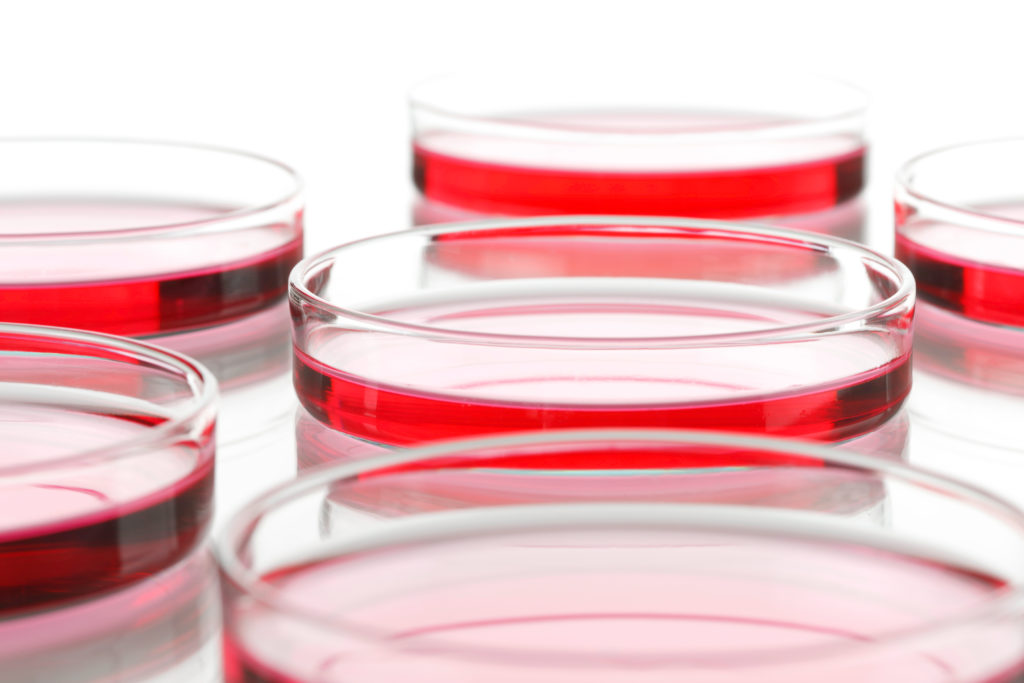
Small blood samples — and the patient-specific, induced pluripotent stem cells (iPSCs) they enable — turn into valuable research material for understanding congenital cardiovascular disorders, especially when united with modern genome sequencing and editing, animal models and three dimensional tissue growth technologies.
Patient-derived induced pluripotent stem cells (iPSCs) have a significant downside for studying most forms of adult heart disease: they resemble fetal heart cells in form and function. But this limitation, which keeps the cells from being easily applicable for the study of many adult conditions, is actually ideal for studying congenital heart problems.
Congenital heart defects or disease (CHD) affect about 1% of all live births in the United States and rank among the leading causes of birth defect-related deaths. These defects can range from mild to very severe, and many require ongoing monitoring and multiple surgeries.
While operations and diagnostics have improved survival for many CHD patients — extending life into adulthood for many children who previously would have died in infancy or early childhood — much remains to be done to advance treatment options and maintenance therapeutics.
Now that patient-derived iPSCs have entered the scene, however, researchers and clinicians are starting to use an exciting term when they discuss the trajectory of current CHD efforts: cure.
iPSCs are excellent for studying CHD, because we can identify potential causative mutations using genome sequencing, then use the cardiomyocytes to look both at the disease and the defects that cause it — in a dish, using just cells, and without a biopsy.
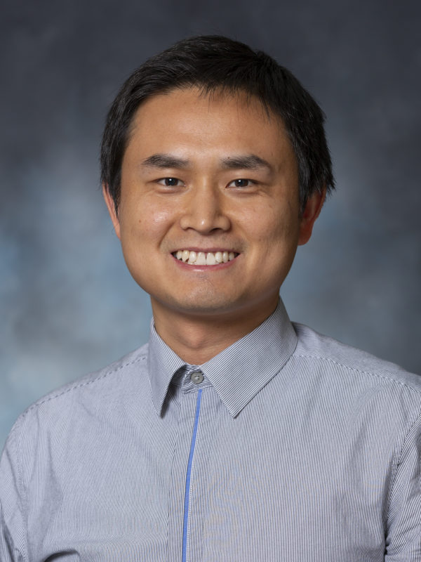
– Mingtao Zhao, DVM, PhD, principal investigator in the Center for Cardiovascular Research in the Abigail Wexner Research Institute at Nationwide Children’s Hospital
Making Cell Magic
Starting with about a teaspoon of blood, researchers separate out peripheral blood mononuclear cells, which they then reprogram into pluripotent stem cells using viral vectors. These are then subjected to a series of treatments to promote differentiation into the primary type of cells found in the heart — cardiomyocytes.
Once this is done properly and the cells ‘beat’ and signal like human heart cells, they can be reproduced so that there are enough to study in various ways: genetic profiling, genome editing, electrophysiological testing, organoid growth (3D tissue generation with multiple cell types), transplantation into animal models, drug screening and more.
“iPSCs are excellent for studying CHD, because we can identify potential causative mutations using genome sequencing, then use the cardiomyocytes to look both at the disease and the defects that cause it — in a dish, using just cells, and without a biopsy,” says Mingtao Zhao, DVM, PhD, principal investigator in the Center for Cardiovascular Research in the Abigail Wexner Research Institute at Nationwide Children’s Hospital. He recently published a protocol for cultivating human cardiomyocytes from routine blood samples; protocols are available for several other heart cell types as well.
Researchers often perform genome or exome screening to identify mutations that are worth studying in greater depth, then they edit the genome of human cardiomyocytes to see if the function of those cells changes.
“Once you know what gene causes what phenotype, you can establish a system to model this disease to get more information,” says Dr. Zhao. “You can find similar mutations in mice or pigs to do further studies on orthologous variants in animals, or use genetic engineering to put the mutation into animals and see if heart defects develop.”
The next step in maximizing the potential of iPSCs for CHD, Dr. Zhao says, will be to correlate the data from iPSC, organoid and animal models to create a more comprehensive picture of the genetic basis of single ventricle heart defects, one of the most severe types of CHD, and — if the impact of the genetic mutation is confirmed — to seek out therapeutic options, such as gene therapy to stimulate the growth of an underdeveloped heart chamber.
Such a dramatic clinical translation of this technology would make a big difference for infants with single ventricle heart disease or an under-developed ventricular chamber, even if it only enlarges the defective heart enough to make surgical interventions more effective.
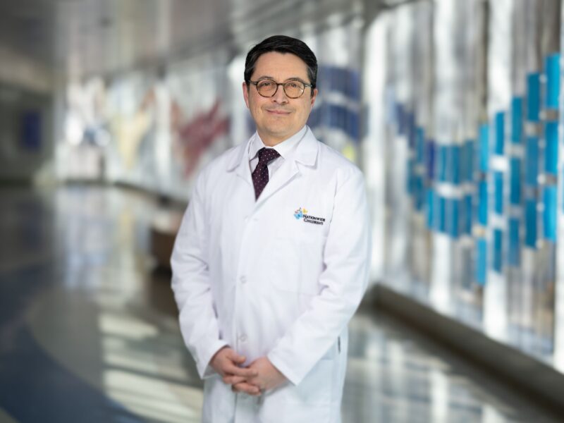
“With hypoplastic left heart syndrome and single ventricle syndromes, our options are palliative — we’re not curing it, and there are major limitations to mouse models for studying therapeutics,” says Vidu Garg, MD, director of the Center for Cardiovascular Research at Nationwide Children’s. Dr. Garg’s research primarily involves murine modeling of CHD and, now, use of iPSCs to help inform tactics taken in his lab.
That’s one of the reasons Dr. Garg recruited Dr. Zhao, who joined Nationwide Children’s in 2019 and has numerous iPSC-related projects now underway. During his time at Stanford University, Dr. Zhao collaborated with iPSC experts such as Joseph Wu, MD, PhD, director of the Stanford Cardiovascular Institute and professor of medicine and radiology at Stanford School of Medicine, who shares Dr. Zhao’s and Dr. Garg’s enthusiasm for the potential these cells hold for cardiovascular research.
“Patient-specific iPSCs are a game changer,” says Dr. Wu, whose lab focuses on iPSCs in cardiac applications, including adult congenital heart disease. “The technology really allows you to understand the first step you need to know to make progress: disease pathology. Then with that you can understand the disease’s mechanism of action, which allows you to look for new drugs and find new therapies, and in turn improve patient care and outcomes.”
Applying the Technology
Isabelle Deschênes, PhD, professor and chair of the Department of Physiology and Cell Biology at The Ohio State University, has studied a large family with congenital heart arrythmias (congenital long QT syndrome, or LQTS) for more than two decades. When iPSCs came on the scene, she was able for the first time to investigate theories of variable genetic penetrance to determine what influences the severity of this potentially fatal — and highly unpredictable — congenital disease.
Her LQTS research showed that iPSCs derived from severely affected family members differed from mildly affected individuals physiologically. Then her team used whole exome sequencing to identify genetic variants present in these patients. Finally, they employed genome editing techniques (using CRISPR genetic engineering) to experiment with the removal or addition of these variants in iPSCs, confirming the roles of multiple genetic modifiers for congenital LQTS.
“Obviously, iPSC cardiomyocytes are a great advance in science, but that’s partly because of great progress in genetics and genomics,” says Dr. Deschênes. “Now, if you suspect that a certain variant in a gene is worsening the phenotype of an individual, you can do CRISPR genome editing to correct it and test the patient-derived cardiomyocytes again. If you see the phenotype is then normal, that proves without a doubt this is why you have that phenotype in that individual.”
The identified protective and aggravating genetic variants were confirmed by the observed phenotypic differences in affected family members when Dr. Deschênes’ team screened the family for the genetic modifiers; the results were published in the Journal of Clinical Investigation.
Dr. Deschênes also collaborates with Dr. Zhao, Dr. Garg and others to advance cardiomyocyte iPSC protocols and applications via her expertise in electrophysiological testing, confirming that the cells function as true heart cells in terms of ion channel activity and electrophysiologic signaling. This allows Drs. Zhao and Garg to use iPSC-derived cardiomyocytes to study the genetics and functional development of single ventricle heart diseases much as Dr. Deschênes did for LQTS patients: identifying potential genetic drivers of single ventricle disease, conducting genome editing, and seeing how the changes affect cell function.
“Making a genetically modified mouse to test some of the variants we can find through exome or genome screening of affected children is possible, but it takes a long time and is a very costly analysis,” says Dr. Garg. “That’s where iPSC technology has added value. Now, we’re taking human cells as opposed to mouse cells, and if we find an interesting genetic variant, we can fix it with genome editing and say, ‘Did it rescue that abnormality?’ There are exciting things we can potentially do with this tool.”
Patient-specific iPSCs are a game changer. The technology really allows you to understand the first step you need to know to make progress: disease pathology. Then with that you can understand the disease’s mechanism of action, which allows you to look for new drugs and find new therapies, and in turn improve patient care and outcomes.
– Joseph Wu, MD, PhD, director of the Stanford Cardiovascular Institute and professor of medicine and radiology at Stanford School of Medicine
The idea may sound fantastic, but it is similar to work already being done on the left side of the heart, for children with hypoplastic left heart syndrome: the abnormal aortic valve is dilated with the hope that it will stimulate growth in the left ventricle by allowing for increased blood flow. iPSCs offer a new avenue to explore such therapeutic options in a host of other single ventricle defects.
Putting iPSCs to Work for a CHD-Free Future
Because the fetal heart is fairly plastic and capable of some remodeling after surgery, Dr. Garg and others think iPSC-driven technology may hold particular promise for early CHD intervention — maybe even a cure.
“With CHD, we’re trying to look at whether iPSC-derived cardiomyocytes from children with single ventricle heart defects respond abnormally to biomechanical forces such as flow and stretch. If we are able to dissect out those mechanisms, it creates the possibility that we could molecularly direct growth or the right biomechanical forces to stimulate cardiomyocytes down their path to proliferate or differentiate,” says Dr. Garg. “So theoretically, we could have a molecular, probably fetal intervention to help a new ventricle grow better in children with single ventricle heart defects. iPSC cardiomyocytes open that door, and could offer a way to almost bypass the primary problem of a valve that isn’t growing.”
Drs. Zhao and Garg have two research endeavors underway applying iPSC technology to a single ventricle heart defect known as pulmonary atresia with intact ventricular septum (PA-IVS). The studies are funded by Additional Ventures, a nonprofit organization that provides significant grant money to advance the science and medicine around single ventricle heart disease.
In one project, they will examine maternal blood for possible cell-free biomarkers for PA-IVS by using patient-derived iPSCs and exosome profiling. The goal would be to predict fetal heart growth and health via a simple blood test for pregnant women.
In their other study, the team will examine patient-specific cardiomyocytes to understand the mechanisms involved in the development of PA-IVS, and whether those mechanisms offer insights for reversing defective heart growth.
“One of the fascinating things you can do is to model what a patient’s outcome was,” says Dr. Garg. “We’re going to have cells from PA-IVS patients, some of whom had bi-ventricle repairs, some of whom followed a single-ventricle pathway. In theory, if the outcomes and response to hemodynamic forces are a genetic phenomenon, we should be able to see differences in terms of ventricular growth by looking at their iPSCs.”
After learning the genetic architecture of single ventricle heart defects such as PA-IVS, investigators may be able to manipulate ventricle growth in animal and 3-D cell models, such as organoids that can form heart chamber-like structures, to help move these advances into formal preclinical therapeutic development. This could eventually lead to a fetal or postnatal intervention to change outcomes for children, from one ventricle to two.
So theoretically, we could have a molecular, probably fetal intervention to help a new ventricle grow better in children with single ventricle heart defects. iPSC cardiomyocytes open that door, and could offer a way to almost bypass the primary problem of a valve that isn’t growing.
– Vidu Garg, MD, director of the Center for Cardiovascular Research in the Abigail Wexner Research Institute at Nationwide Children’s Hospital
The science will take time, though, and translation to the clinic will be complex.
“Cell culture models are not living animal models, and CHD involves multiple cells, so how do you know what you’re seeing in one cell type is truly what is happening in patients? That’s one issue,” says Dr. Garg. “There are weaknesses to the mouse model as well — it can’t serve as the CHD model when studying CHD-associated long-term morbidities. We need to create better models of disease if we’re going to come up with therapeutics or novel prevention strategies. That’s where cardiac organoid and 3-D tissue differentiation are going to improve things and offer a lot of advantages for bringing these findings to the clinic.”
Making Precision Medicine Possible
“We often joke that we’ve cured heart disease a million times over in mice, but it hasn’t always translated over to humans,” says Dr. Deschênes. “Working with human cells is a great advantage. iPSCs are not perfect, but they are one of the best options we have right now. The beauty is that they’re a patient’s real cells, not a model, so we can see what’s happening in that patient — what’s driving their disease and what are the potential targets we can hit to improve their health.”
Dr. Zhao, who recently published a paper describing the potential synergy across research modalities for CHD, believes that a combination of iPSC, genomic, animal, and organoid research methodologies could also carry other areas of medicine to new heights.
“Single cell genomics from iPSCs can give us very useful information regarding many other disease mechanisms that could allow personalized medicines and treatments, even beyond a gene therapy to grow a new heart chamber,” he says.
In that vein, Dr. Wu sees enormous potential for iPSCs to enable “clinical trials in a dish” — rapid screening for potential adverse effects or efficacy on a large number of patients (through their cell lines) prior to making the investment in a true clinical trial. To aid in this and other research efforts, he has built the largest academic bank of iPSCs in the country, with more than 1500 patient samples.
Rather than using animal studies that typically involve clones — wherein testing on 1,000 mice is essentially testing just one mouse and 999 identical twins likely to be affected the same way — the iPSC bank could enable rapid screening for potential benefits or risks of a drug for 1,000 genetically different individuals. A 50-50 sex ratio and customizable ethnicity breakdown could allow valuable preclinical insight regarding who might be good responders, and thus who would be best to include in a clinical trial for that compound.
“Once you have the ‘clinical trial in a dish’ model, fast forward 10-20 years, and it should be possible to have true precision medicine,” says Dr. Wu, who has already demonstrated the potential for this approach with iPSCs and LMNA-related dilated cardiomyopathy, published in Science Translational Medicine. “For example, if you take a drug, how do you know if it will really work for you or not? In the future, we will use iPSCs and genetic screening to help understand who will improve and who won’t. In my opinion that’s the holy grail of precision medicine.”
Now, if you suspect that a certain variant in a gene is worsening the phenotype of an individual, you can do CRISPR genome editing to correct it and test the patient-derived cardiomyocytes again. If you see the phenotype is then normal, that proves without a doubt this is why you have that phenotype in that individual.
– Isabelle Deschênes, PhD, professor and chair of the Department of Physiology and Cell Biology at The Ohio State University
The key to precision medicine and a CHD cure alike will be translation of the power of iPSCs and complementary technologies into the clinic.
“I often think some of these innovations are the answer we’ve been looking for — that we’re finally going to do it! But can we really move the knowledge we’ve gained into clinical practice?” asks Dr. Garg. “Time will tell. I think we can.”
This article appears in the 2021 Fall/Winter print issue. Download the full issue.
References:
- Chai S, Wan X, Ramirez-Navarro A, Tesar PJ, Kaufman ES, Ficker E, George AL Jr, Deschênes I. Physiological genomics identifies genetic modifiers of long QT syndrome type 2 severity. J Clin Invest. 2018 Mar 1;128(3):1043-1056.
- Lin H, McBride KL, Garg V, Zhao MT. Decoding genetics of congenital heart disease using patient-derived induced pluripotent stem cells (iPSCs). Front Cell Dev Biol. 2021 Jan 21;9:630069.
- Sayed N, Liu C, Ameen M, Himmati F, Zhang JZ, Khanamiri S, Moonen JR, Wnorowski A, Cheng L, Rhee JW, Gaddam S, Wang KC, Sallam K, Boyd JH, Woo YJ, Rabinovitch M, Wu JC. Clinical trial in a dish using iPSCs shows lovastatin improves endothelial dysfunction and cellular cross-talk in LMNA cardiomyopathy. Sci Transl Med. 2020 Jul 29;12(554):eaax9276.
- Ye S, Wan X, Su J, Patel A, Justis B, Deschênes I, Zhao MT. Generation and expansion of human cardiomyocytes from patient peripheral blood mononuclear cells. J Vis Exp. 2021 Feb 12;(168).
Image credits: iStock (header); Nationwide Children’s (portraits)
In 2020, Nationwide Children’s received a $1 million Innovation Fund endowed by Additional Ventures, joining a handful of other research institutions in a large-scale coordinated effort to find new ways to functionally cure patients with SV CHD. To learn more about this funding, read Additional Ventures: Investing in Congenital Heart Disease Research to Advance Care.
About the author
Katherine (Katie) Brind’Amour is a freelance medical and health science writer based in Pennsylvania. She has written about nearly every therapeutic area for patients, doctors and the general public. Dr. Brind’Amour specializes in health literacy and patient education. She completed her BS and MS degrees in Biology at Arizona State University and her PhD in Health Services Management and Policy at The Ohio State University. She is a Certified Health Education Specialist and is interested in health promotion via health programs and the communication of medical information.
- Katie Brind'Amour, PhD, MS, CHEShttps://pediatricsnationwide.org/author/katie-brindamour-phd-ms-ches/

- Katie Brind'Amour, PhD, MS, CHEShttps://pediatricsnationwide.org/author/katie-brindamour-phd-ms-ches/
- Katie Brind'Amour, PhD, MS, CHEShttps://pediatricsnationwide.org/author/katie-brindamour-phd-ms-ches/
- Katie Brind'Amour, PhD, MS, CHEShttps://pediatricsnationwide.org/author/katie-brindamour-phd-ms-ches/