InSight: Working Up the Nerve
InSight: Working Up the Nerve https://pediatricsnationwide.org/wp-content/themes/corpus/images/empty/thumbnail.jpg 150 150 Katie Brind'Amour, PhD, MS, CHES https://pediatricsnationwide.org/wp-content/uploads/2021/03/Katie-B-portrait.gifUsing regional anesthesia to numb nerves reduces pain and speeds recovery in pediatric orthopedic surgery.
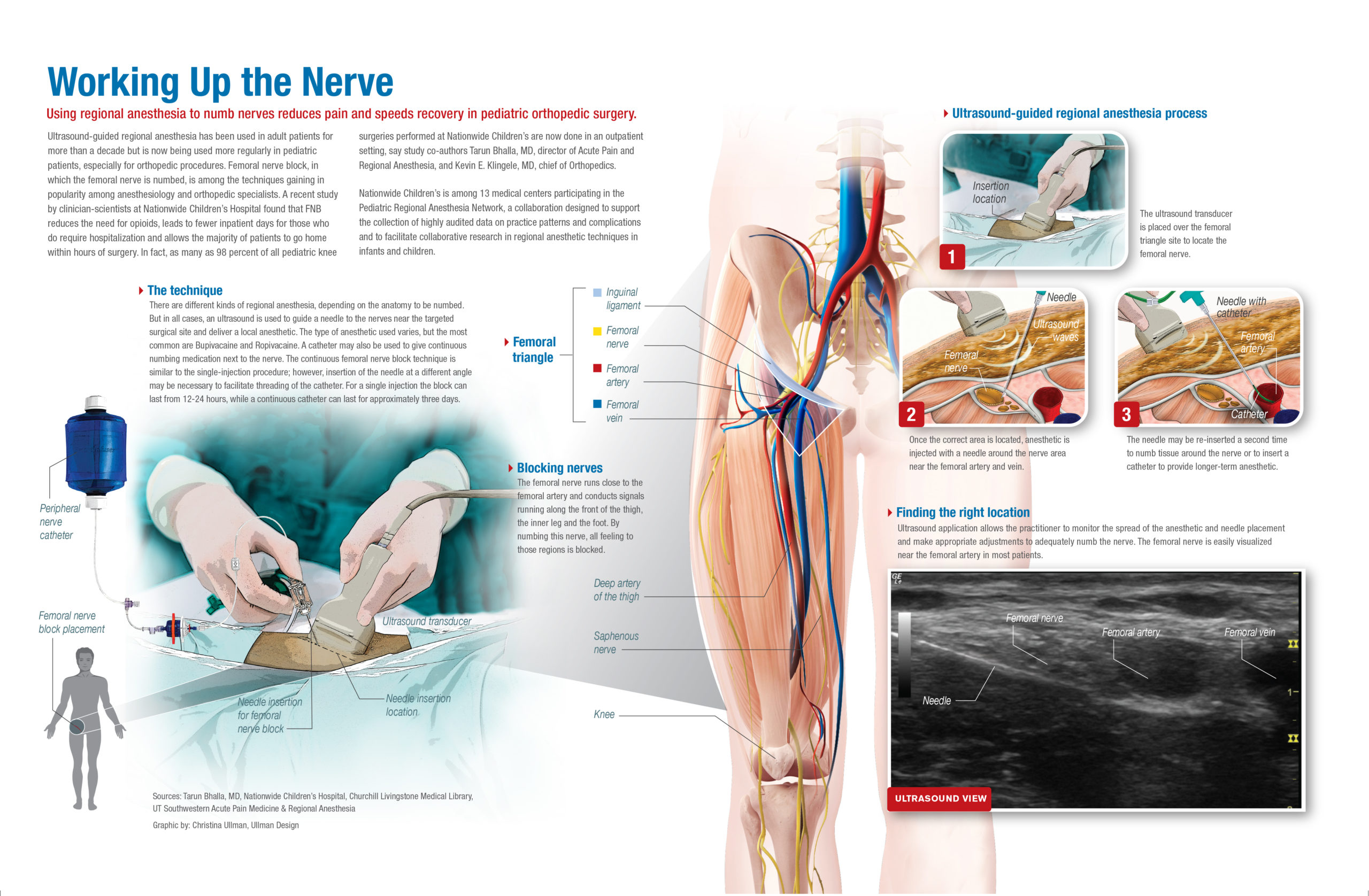
Ultrasound-guided regional anesthesia has been used in adult patients for more than a decade but is now being used more regularly in pediatric patients, especially for orthopedic procedures. Femoral nerve block, in which the femoral nerve is numbed, is among the techniques gaining in popularity among anesthesiology and orthopedic specialists.
A recent study by clinician-scientists at Nationwide Children’s Hospital found that FNB reduces the need for opioids, leads to fewer inpatient days for those who do require hospitalization and allows the majority of patients to go home within hours of surgery. In fact, as many as 98 percent of all pediatric knee surgeries performed at Nationwide Children’s are now done in an outpatient setting, say study co-authors Tarun Bhalla, MD, director of Acute Pain and Regional Anesthesia, and Kevin E. Klingele, MD, chief of Orthopedics.
Nationwide Children’s is among 13 medical centers participating in the Pediatric Regional Anesthesia Network, a collaboration designed to support the collection of highly audited data on practice patterns and complications and to facilitate collaborative research in regional anesthetic techniques in infants and children.
The Technique
There are different kinds of regional anesthesia, depending on the anatomy to be numbed. But in all cases, an ultrasound is used to guide a needle to the nerves near the targeted surgical site and deliver a local anesthetic. The type of anesthetic used varies, but the most common are Bupivacaine and Ropivacaine. A catheter may also be used to give continuous numbing medication next to the nerve. The continuous femoral nerve block technique is similar to the single-injection procedure; however, insertion of the needle at a different angle may be necessary to facilitate threading of the catheter. For a single injection, the block can last from 12-24 hours, while a continuous catheter can last for approximately three days.
The femoral nerve runs close to the femoral artery and conducts signals running along the front of the thigh, the inner leg and the foot. By numbing this nerve, all feeling to those regions is blocked.
First, the ultrasound transducer is placed over the femoral triangle site to locate the femoral nerve. Next, the anesthetic is injected with a needle around the nerve area near the femoral artery and vein. Finally, the needle may be re-inserted a second time to numb tissue around the nerve or to insert a catheter to provide longer-term anesthetic.
Ultrasound application allows the practitioner to monitor the spread of anesthetic and needle placement and make appropriate adjustments to adequately numb the nerve. The femoral nerve is easily visualized near the femoral artery in most patients.
This article appeared in the Fall/Winter 2014 print issue. Download the full issue now.
About the author

Katherine (Katie) Brind’Amour is a freelance medical and health science writer based in Pennsylvania. She has written about nearly every therapeutic area for patients, doctors and the general public. Dr. Brind’Amour specializes in health literacy and patient education. She completed her BS and MS degrees in Biology at Arizona State University and her PhD in Health Services Management and Policy at The Ohio State University. She is a Certified Health Education Specialist and is interested in health promotion via health programs and the communication of medical information.
- Katie Brind'Amour, PhD, MS, CHEShttps://pediatricsnationwide.org/author/katie-brindamour-phd-ms-ches/

- Katie Brind'Amour, PhD, MS, CHEShttps://pediatricsnationwide.org/author/katie-brindamour-phd-ms-ches/
- Katie Brind'Amour, PhD, MS, CHEShttps://pediatricsnationwide.org/author/katie-brindamour-phd-ms-ches/
- Katie Brind'Amour, PhD, MS, CHEShttps://pediatricsnationwide.org/author/katie-brindamour-phd-ms-ches/