
New Approach to Dystrophin Quantification Can Help Researchers Studying Muscular Dystrophy
New Approach to Dystrophin Quantification Can Help Researchers Studying Muscular Dystrophy https://pediatricsnationwide.org/wp-content/themes/corpus/images/empty/thumbnail.jpg 150 150 Abbie Miller https://pediatricsnationwide.org/wp-content/uploads/2023/05/051023BT016-Abbie-Crop.jpgDystrophin is the protein product of the DMD gene and plays an important role in Becker and Duchenne muscular dystrophies.
Researchers in the Center for Gene Therapy at the Abigail Wexner Research Institute at Nationwide Children’s Hospital have developed an automated, unbiased approach for the precise quantification of dystrophin immunofluorescence in muscle samples.
Dystrophin is the protein product of the DMD gene, and it has an important role in muscle integrity. The DMD gene is large and vulnerable to many different mutations that can affect the amount and quality of protein produced. These mutations lead to the neuromuscular diseases we know as Becker or Duchenne muscular dystrophies. Even among these two forms of DMD-mediated muscular dystrophy, a variety of phenotypes are possible.
“A fundamental question in research related to DMD-mediated muscular dystrophies is how much dystrophin is enough, or more specifically, how does the level of dystrophin present correlate with the range of mutations and phenotypes we see in the clinic?” says Kevin Flanigan, MD, director of the Center for Gene Therapy. “And when it comes to restorative therapies, those aimed at replacing the dystrophin gene, how much dystrophin is enough to make a therapy effective?”
Answering these questions requires researchers to have a consistent, reliable and unbiased way to quantify the amount of dystrophin in a sample. Numerous methods exist, but they’re often expensive.
“We wanted to create something accessible – that is, easy to use and affordable for a variety of labs doing muscular dystrophy research,” says Tatyana Vetter, PhD, research scientist in the Center for Gene Therapy and director of Microscopic Imaging for the center.


Using microscope images of whole-tissue sections stained for dystrophin and spectrin, Dr. Vetter developed a method, built on a commonly used microscopy software platform, that can assess the dystrophin density and proportion of dystrophin-positive coverage at the sarcolemma of each muscle fiber. To safeguard objectivity, she used thresholds for dystrophin and spectrin that are derived empirically from non-sarcolemmal signal intensity within each tissue section.
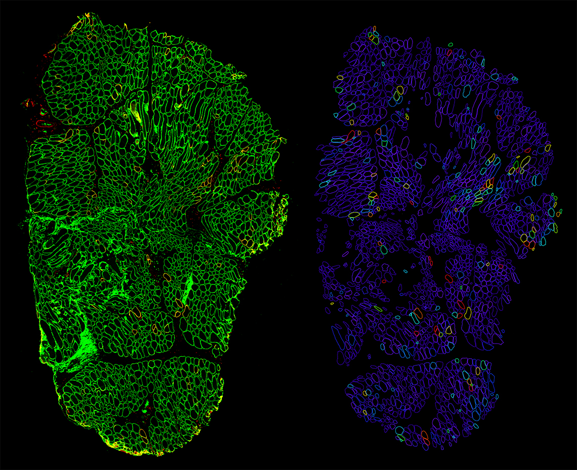
(Left) Original IF microscopy of muscle fiber showing spectrin in green and dystrophin in red. (Right) Positivity analysis output.
“In developing and validating the program, I ensured I would be able to train any user to obtain repeatable and reliable measurements,” Dr. Vetter says. “By creating what the software calls ‘analysis recipes,’ we can make the learning curve very small. Users don’t have to know how to write code or tinker with the analysis algorithm to get the measurements they need.”
Looking forward, Drs. Vetter and Flanigan have already looked at how the methodology can be used to measure other proteins in tissue samples.
“This technique is not limited to dystrophin – or even to muscle fibers,” says Dr. Flanigan. “We are already exploring how to use this tool to measure important proteins in other neuromuscular conditions, and we’re collaborating with other labs, and even other disciplines.”
The methodology is available free of charge for researchers. Interested labs should contact the Center for Gene Therapy.
Reference:
Vetter TA, Nicolau S, Bradley AJ, Frair EC, Flanigan KM. Automated immunofluorescence analysis for sensitive and precise dystrophin quantification in muscle biopsies. Neuropathology and Applied Neurobiology. 2021 Nov 30. [Epub ahead of print]
Image credit: Nationwide Children’s Hospital
About the author

Abbie (Roth) Miller, MS, MWC, is a passionate communicator of science. As the manager of medical and science content at Nationwide Children’s Hospital, she shares stories about innovative research and discovery with audiences ranging from parents to preeminent researchers and leaders. She is a Medical Writer Certified®, credentialed by the American Medical Writers Association, and received her masters of science in Health Communication from Boston University.
- Abbie Millerhttps://pediatricsnationwide.org/author/abbie-miller/

- Abbie Millerhttps://pediatricsnationwide.org/author/abbie-miller/
- Abbie Millerhttps://pediatricsnationwide.org/author/abbie-miller/
- Abbie Millerhttps://pediatricsnationwide.org/author/abbie-miller/