Evolution of an Atlas
Evolution of an Atlas https://pediatricsnationwide.org/wp-content/themes/corpus/images/empty/thumbnail.jpg 150 150 Katie Brind'Amour, PhD, MS, CHES Katie Brind'Amour, PhD, MS, CHES https://pediatricsnationwide.org/wp-content/uploads/2021/03/Katie-B-portrait.gif- April 28, 2014
- Katie Brind'Amour, PhD, MS, CHES
Adult brain atlases have existed for years. Why is it so crucial — and so difficult — to build one for preemies?
Even experts need maps. They give perspective, scale and orientation. They can show both current location and the final destination. And in the world of premature brains, they can offer vital information about subtle injury, developmental delay and opportunities for intervention.
But existing brain maps primarily feature adult brains, which have different landmarks. Newer maps of healthy, full-term infant brains have different scales and features than their less-developed preemie counterparts. So, how do you go about building a map for the premature brain?
This is a challenge neonatologist Nehal Parikh, DO, MS, has tackled at Nationwide Children’s Hospital, taking brain mapping into uncharted territory. When he set out to develop an atlas of the premature brain, he first needed to overcome two not-so-simple problems: the subjectivity of MRI diagnostics and an entire profession’s lack of knowledge about brain development in premature babies.
ATLAS HISTORY
Clinician researchers started applying mapping techniques to aid our understanding of the human brain more than 150 years ago. Each map featured a single individual’s characteristics in 2-D illustrations, drawn from cadaver brains. The most widely referenced series of paper brain maps, created in 1909 by German anatomist Korbinian Brodmann, guided surgeons and pathologists in their craft late into the 20th century.
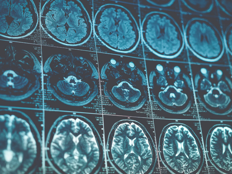
Now, adult brain maps are organized into digital atlases that resemble a car’s GPS in their level of sophistication — they can embed layers of information to tell users about brain segment density, volume, blood flow and genetics much like a car’s computer can identify local restaurants and estimate travel time based on current speed. The most advanced adult brain atlases include population-based templates built by combining scans from hundreds of individuals to figure out what normal parameters are for each brain feature.
“In adults, probabilistic, population-based brain atlases provide an assessment of the normal brain at different ages,” says John Mazziotta, MD, PhD, executive vice dean of the David Geffen School of Medicine at the University of California, Los Angeles and father of the modern adult brain atlas. “Since the normal human brain varies in size, shape and configuration, having an average brain atlas provides a basis for determining subtle abnormalities, as these fall outside the range of normal variance.”
Dr. Parikh has spent the past 10 years trying to achieve the same level of complexity in premature infant brain imaging. His first attempts to reduce the subjectivity of MRI interpretation, begun during his time in Houston at the University of Texas, involved an algorithm for manual, objective evaluation of neonatal MRI scans that appeared to indicate subtle injuries to the brain’s white matter.
“I failed miserably,” he admits. “But it did help me understand why radiologists weren’t already objectively defining these injuries in preemies.”
His techniques for researching the anatomy and abnormalities of the premature brain have since evolved to include digital automation processes. By starting with adult brain segmentation computer software developed by Ponnada Narayana, PhD, at The University of Texas Medical School at Houston, Dr. Parikh helped develop automated brain segmentation software for preterm infants. The technique required years of focused work to first reliably and reproducibly segment developing brain tissues and structures that lacked clear anatomic boundaries.
His new measures resulted in what would become the first layer of his atlas: one that allows the automatic, objective quantification of brain tissues and subtle but diffuse injuries not previously measured by traditional diagnostic imaging technology. Having removed some of the subjectivity from the MRI diagnostic process, Dr. Parikh, also a principal investigator in the Center for Perinatal Research in The Research Institute at Nationwide Children’s, then turned his attention to the creation of a multi-layer, digital atlas of the premature brain.
PEERING INTO THE PREMATURE BRAIN
Dr. Parikh’s mission hinged on one task. He would have to study hundreds of MRI scans of preemies to define what the average premature brain actually looks like. These infant brains are obviously much smaller than those of adults, but they also contain more water, more immature structures and fewer connective networks.
To get from a series of scans to an actual template for an atlas, Dr. Parikh applied the same principle to mapping the premature brain as Dr. Mazziotta used to build his adult brain atlas. Numerous subjects’ brain images were compared, manually segmented, measured for key parameters and averaged to obtain normal ranges. Ideally, a new brain image could then be contrasted with the population-based template to visually and mathematically identify abnormalities.
“I spent a lot of time in the early years working with neuroradiologists and physicists, sending out emails to people I didn’t know to see if they would share their neonatal imaging sequences with us to tailor them for our premature population,” Dr. Parikh recalls. “The question is, how do you come up with the best atlas for such a unique group? I think that’s an evolving issue as we continue to build on prior work.”
Very little is known about the appearance of a truly normal premature brain. What is the difference between underdevelopment due to prematurity versus that due to injury? What is healthy for a preemie brain, especially if “healthy” isn’t average?
To further complicate the matter, the premature brain doubles in size between 28 and 40 weeks postmenstrual age, literally making a brain atlas for this population a moving target.
“Right now, we’re creating atlases with and without subtle injury, trying to see which one works out best in the end with outcome measures,” Dr. Parikh says.
ATLAS-MAKING IN ACTION
Despite the simultaneous efforts of a few teams around the world, the premature brain atlas is definitely a work in progress.
One way to tackle the challenge of building a useful atlas, Dr. Parikh explains, is to follow premature babies over time for functional outcome measures that correlate with the individual’s brain images in infancy. This would tell researchers whether certain brain injuries and underdeveloped segments during infancy actually impact long-term development. A series of images on the same children combined with functional outcome measures could offer the precision needed, Dr. Parikh says.
A second option, he suggests, is to build atlases constructed with images of premature infants at 28, 30, 32 and 40 weeks postmenstrual age and 3 months corrected age. This could provide a more appropriate picture of a developmental trajectory — with true clinical implications, Dr. Parikh says. His team is pursuing both options.
“If a baby’s development falls off that trajectory, that may be a good predictor of delays and impairments down the road,” he says. “It could also serve as an early indicator that this baby might benefit from intervention, instead of waiting two or more years for problems to appear.”
This conviction comes from conveying his fragile patients in a special MRI-compatible incubator to the radiology department hundreds of times. Dr. Parikh uses their results to refine the science and to improve his clinical suggestions to anxious parents eager to have any advanced notice of potential developmental difficulties in their premature babies.
MAPPING MOTIVATION
As a clinician, effective therapeutic intervention and improved long-term outcomes are Dr. Parikh’s ultimate aims. As a clinical trialist, more efficient research and faster translation of results are his goals.
His probabilistic atlas already enables more objective, accurate diagnoses and increased sensitivity in the detection of subtle brain injury. But by embedding as much relevant information as possible into the program, the tool could have much wider use in neonatology. EEG data, brain tract volumes, functional MRI measures, blood flow, genetic information, clinical risk factors, metabolite measures and neuron networking could all be added to his current atlas to improve the tool’s diagnostic and predictive power.
“I want to be able to offer an accurate prognosis and better strategies to prevent the neurodevelopmental disabilities that affect 40 percent of these preemies,” Dr. Parikh explains. “An atlas like this would have tremendous potential for predicting clinical outcomes and for enabling a new model for faster, more efficient research.”
A wide range of embedded features could help measure intervention effectiveness within weeks or months instead of years. Therapies that aren’t working can be quickly discontinued and substituted with another intervention.
“Until we develop that robust, multi-modal program, we can still work with the individual layers of that atlas to inform our clinical decision-making and research,” Dr. Parikh says. “But the days of using automated MRI atlas diagnostics in routine clinical care of premature babies may be only five or 10 years away.”
About the author
Katherine (Katie) Brind’Amour is a freelance medical and health science writer based in Pennsylvania. She has written about nearly every therapeutic area for patients, doctors and the general public. Dr. Brind’Amour specializes in health literacy and patient education. She completed her BS and MS degrees in Biology at Arizona State University and her PhD in Health Services Management and Policy at The Ohio State University. She is a Certified Health Education Specialist and is interested in health promotion via health programs and the communication of medical information.
-
Katie Brind'Amour, PhD, MS, CHEShttps://pediatricsnationwide.org/author/katie-brindamour-phd-ms-ches/April 27, 2014
-
Katie Brind'Amour, PhD, MS, CHEShttps://pediatricsnationwide.org/author/katie-brindamour-phd-ms-ches/April 27, 2014
-
Katie Brind'Amour, PhD, MS, CHEShttps://pediatricsnationwide.org/author/katie-brindamour-phd-ms-ches/April 27, 2014
-
Katie Brind'Amour, PhD, MS, CHEShttps://pediatricsnationwide.org/author/katie-brindamour-phd-ms-ches/April 28, 2014
- Post Tags:
- Neonatology
- Neurology
- Posted In:
- Features


