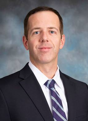
Featured Researcher — Mitchel Stacy, PhD
Featured Researcher — Mitchel Stacy, PhD https://pediatricsnationwide.org/wp-content/themes/corpus/images/empty/thumbnail.jpg 150 150 Katie Brind'Amour, PhD, MS, CHES https://pediatricsnationwide.org/wp-content/uploads/2021/03/Katie-B-portrait.gifMitchel Stacy, PhD, a principal investigator in the Center for Regenerative Medicine at Nationwide Children’s Hospital and assistant professor of Surgery at The Ohio State University, started his education with dietetics in mind. Finding his way instead to nuclear and molecular imaging of cardiovascular diseases, Dr. Stacy’s research blossomed during his postdoctoral fellowship and first faculty position at Yale University.
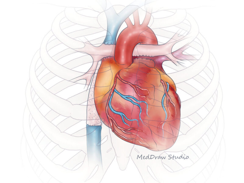
There, he began studying the ability of nuclear imaging to assess adults with cardiovascular disease and evaluate remodeling of tissue-engineered vascular grafts designed and placed by Toshiharu Shinoka, MD, PhD, and Christopher Breuer, MD, co-directors of the Center for Regenerative Medicine at Nationwide Children’s.
Since joining Nationwide Children’s in 2018, Dr. Stacy has reinstituted his work with Drs. Breuer and Shinoka and added numerous other clinicians and researchers to his list of collaborators.
Dr. Stacy focuses on the advancement of nuclear and multimodality imaging techniques, such as computed tomography (CT) angiography, single photon emission computed tomography (SPECT)/CT, positron emission tomography (PET)/CT, and ultrasound. He is especially interested in their utility in non-invasively assessing tissue remodeling and disease status, prognosis and progression for a wide variety of clinical conditions. By studying imaging from patients and preclinical models, Dr. Stacy and his team can identify key indicators of cardiovascular events or remodeling of tissue-engineered scaffolds, for example.
Dr. Stacy is currently working to expand his investigations from adult cardiovascular imaging to imaging of pediatric cardiology and oncology. A recent study by Dr. Stacy’s lab involving retrospective analysis of 10 years’ worth of PET/CT images performed in pediatric oncology patients at Nationwide Children’s revealed the potential of this imaging to predict cardiovascular complications, such as thrombotic events. Dr. Stacy suggests that if this risk could be understood in advance, clinicians could potentially adjust medications to reduce the chance of thrombosis. He recently submitted a National Institutes of Health grant proposal that would involve expanding on this work and prospectively following about 400 children with lymphoma from numerous pediatric hospitals in the Midwest. The focus of this multi-site investigation is to further evaluate the possible connection between his PET/CT imaging model and risk for thrombosis in this population.
This type of direct translational impact is at the heart of Dr. Stacy’s research program — learning from distinct biological and physiological clues visible from multimodal imaging and turning them into meaningful clinical predictions and insights.
Read on to learn more about Dr. Stacy and his work.
What was your path to your current role?
If someone asked me 20 years ago what I would be doing, I’m not sure I could have predicted where I am now.
Throughout my childhood and into my 20s, I was involved in competitive distance running, which generated a lifelong interest in cardiovascular health and nutrition. I completed my undergraduate degree in dietetics at The Ohio State University and initially thought I would become a dietitian.
Instead, I ultimately decided to pursue a doctorate at the University of Toledo that was focused on exercise and cardiovascular physiology.
During that program, I had a chance to learn about ultrasound, which jumpstarted my interest in everything imaging related and ultimately led me to a postdoctoral fellowship in cardiovascular imaging at Yale School of Medicine. Through my fellowship program, I got exposure to additional imaging methods, including PET/CT, SPECT/CT and MRI, and I learned how to apply these techniques to a variety of different cardiovascular diseases and clinical contexts. I also met and had the opportunity to collaborate with Drs. Breuer and Shinoka, performing imaging studies to evaluate remodeling of their tissue-engineered vascular graft. They moved from Yale to Nationwide Children’s in 2012, and I transitioned into a junior faculty role in the cardiology program at Yale for several years.
In 2018, Dr. Breuer and I had the opportunity to reconnect, and he shared that the Center for Regenerative Medicine at Nationwide Children’s was hiring. He ultimately recruited me to set up a translational imaging-focused research program.
Fun Facts About Dr. Stacy
What’s your favorite word, and why?
“Daddy.” My son is at a fun age where he is beginning to talk, and listening to him call for me is one of the most exciting parts of my day.
What do you usually eat for breakfast?
I keep it pretty simple and usually eat the same thing to save time as I run out the door: a banana, protein bar and multiple coffees throughout the morning.
What’s your dream job, besides working in research?
I would love to be a travel blogger who provides recommendations on where to visit, stay or eat. My wife and I have been to Bali, Bermuda, Italy, Spain, Australia and more, and we’d like to keep exploring — Iceland and New Zealand are on the list.
What’s your favorite food?
I generally like any type of seafood. I could eat tuna, salmon or some form of sushi for every meal.
Favorite way to relax?
I usually exercise in some way every evening, which is the one time of day when I can really shut my brain off and relax. On the weekends, I enjoy spending quality time with my family.
Since my arrival at Nationwide Children’s, we have rejuvenated our imaging-related collaboration in cardiovascular tissue engineering. I now primarily focus on applying non-invasive imaging techniques to a variety of different clinical needs, including tissue engineering, pediatric cardiology, pediatric oncology and adult cardiovascular diseases.
What is your favorite part of your job?
Every day is different, and I’m constantly learning. Some days I’m sitting in my office writing a paper or a grant application, while other days might involve imaging patients, processing medical images, or learning new imaging or surgical techniques. That variety is what I enjoy the most about being involved in research — I never get bored.
I also love sharing the potential of molecular imaging, which is still a relatively new concept in the field of radiology. Much like MRI, molecular imaging with nuclear medicine techniques is an evolving field that is leading to new applications for imaging in the clinical environment. Many people have this perception of nuclear imaging as a scary thing due to radiation exposure, but it can provide new insight into the pathophysiology of different diseases that isn’t possible with other imaging techniques. It’s exciting to collaborate with clinicians and researchers to introduce them to new imaging paradigms. Being able to prove these imaging methods have translatable clinical value is really what makes my work worthwhile.
How does your research serve our patients and our community?
Research in my lab is focused on developing new, non-invasive imaging techniques or capitalizing on existing imaging technology that can allow us to better understand cardiovascular physiology and disease. We also develop new imaging measures to predict how patients will respond to certain treatments and predict future health problems or outcomes. The results of these imaging studies can help guide clinicians with treatment planning, improve evaluation of cardiovascular diseases, reduce medical costs and ultimately improve patient outcomes.
For example, in my work with Drs. Breuer and Shinoka, we are establishing PET/CT imaging as a marker of the inflammatory response to implanted tissue-engineered vascular grafts. By developing a non-invasive method to physiologically evaluate how these biomaterials evolve after implantation, we can start to predict the long-term remodeling characteristics of tissue-engineered vascular grafts that are now used to treat children born with congenital heart disease. Evaluation of inflammation via PET/CT imaging could ultimately assist clinicians with better understanding how these grafts are performing and potentially guide future monitoring or treatment of these patients. Additionally, PET/CT imaging of inflammation may help optimize the design of future vascular grafts composed of different biomaterials that produce a lower inflammatory response in the body.
Similarly, in children with cancer who have undergone treatment with chemotherapy, we can use PET/CT imaging to evaluate the inflammatory response to chemotherapy in their blood vessels, which in turn might predict their risk for future thrombotic events, such as deep vein thrombosis or pulmonary embolism. We expect this will help determine which patients are at highest risk for thrombosis and assist physicians with the timing and tailoring of anticoagulant treatments. Essentially, non-invasive imaging can function as a predictive biomarker that can give clinicians valuable information about how to better treat their patients.
So, what’s next?
Ideally, I would like to establish a leading translational molecular imaging program here at Nationwide Children’s. I also hope to continue developing interdisciplinary imaging collaborations with other research teams at Nationwide Children’s and The Ohio State University.
Imaging is relevant to every type of disease, and many clinical or pre-clinical research studies could probably benefit from incorporating imaging in some way. For example, we recently learned through collaboration with another research team that it is possible to conduct microCT imaging of anesthetized crayfish. This is potentially important because crayfish serve as a good translational model for exploring tissue regeneration. I would like for my imaging-related work to help progress research in a variety of fields and ultimately have a demonstrable impact on patient care and outcomes.
About the author
Katherine (Katie) Brind’Amour is a freelance medical and health science writer based in Pennsylvania. She has written about nearly every therapeutic area for patients, doctors and the general public. Dr. Brind’Amour specializes in health literacy and patient education. She completed her BS and MS degrees in Biology at Arizona State University and her PhD in Health Services Management and Policy at The Ohio State University. She is a Certified Health Education Specialist and is interested in health promotion via health programs and the communication of medical information.
- Katie Brind'Amour, PhD, MS, CHEShttps://pediatricsnationwide.org/author/katie-brindamour-phd-ms-ches/

- Katie Brind'Amour, PhD, MS, CHEShttps://pediatricsnationwide.org/author/katie-brindamour-phd-ms-ches/
- Katie Brind'Amour, PhD, MS, CHEShttps://pediatricsnationwide.org/author/katie-brindamour-phd-ms-ches/
- Katie Brind'Amour, PhD, MS, CHEShttps://pediatricsnationwide.org/author/katie-brindamour-phd-ms-ches/