Distal Radius Physeal Bar and Ulnar Overgrowth: Indications for Treatment
Distal Radius Physeal Bar and Ulnar Overgrowth: Indications for Treatment https://pediatricsnationwide.org/wp-content/uploads/2020/06/122016ds0034-samora-header-1024x575.jpg 1024 575 Julie Samora, MD, PhD Julie Samora, MD, PhD https://secure.gravatar.com/avatar/046f18da46df408fb88883823eeead0d?s=96&d=mm&r=g- June 04, 2020
- Julie Samora, MD, PhD

Distal radius fractures are among the most common fractures in pediatrics. Although most heal without complication, some result in partial or complete physeal arrest. Risk factors for distal radius growth arrest include physeal fractures, ischemia, infection, radiation, tumor, blood dyscrasias, burns, frostbite and repetitive stress. The distal radius physis is responsible for 75% of the longitudinal growth of the radius, with expected closure at approximately 15 years of age in females and 17 years in males. The size, shape and location of the bony bridge as well as the age of the child will dictate the outcomes. Radial growth arrest can lead to angular deformity, abnormal wrist mechanics, ulnar overgrowth and distal radial ulnar joint (DRUJ) instability.
Patients with a growth arrest of the distal radius can present with ulnar-sided wrist pain and sometimes a prominence of the ulnar head. There is occasionally loss of pronosupination of the forearm and radial/ulnar deviation of the wrist. The DRUJ should be assessed for any instability or side-to-side differences.
If growth arrest of the distal radius is suspected, treatment decisions should be made on a variety of factors, considering patient age, remaining growth potential, location of injury within the physis, size of physeal bar, angular deformity, and individual patient and family desires. Categories of treatment options include observation, physeal bar resection, epiphysiodeses, osteotomies and distraction osteogenesis. In cases where minimal growth remains, and if the size of the bar is small, conservative management is the recommended course of action.
Physeal bar resection attempts to completely remove the bar and leave surrounding healthy physis. Indications include bar < 50%, more than 2 years of growth remaining, and angular deformity < 20̊. Identification of the exact location of the bar is necessary to perform this surgery accurately, which requires advanced imaging, such as MRI. Regardless of technique, physeal bar resection is difficult and even if complete resection is obtained, the surrounding physis may be affected and may not respond to the untethering surgery. The results of this surgery are unpredictable and usually even with some growth after the surgery, it is usually not sufficient to catch up with the ulnar growth.
In cases of partial arrest, completion epiphysiodesis can prevent angular deformity that would occur from continued, asymmetric growth. If there is a complete distal radius growth arrest, an ulnar epiphysiodesis should be performed to prevent positive ulnar variance. Epiphysiodesis can be performed in multiple ways, including mini-open approaches, percutaneous drilling, open curettage and open burr resection.
With ulnar positive variance, there is tremendous load on the lunate, triquetrum, and TFCC. An ulnar shortening osteotomy (USO) is a very reliable surgery that can decrease or completely eliminate pain from unequal growth of the ulna and improve wrist mechanics and DRUJ stability. Depending on how much growth is remaining, an USO as well as an ulnar epiphysiodesis must be performed. Results of USO are generally good with decreased pain and improved or similar modified wrist scores.
If an angular deformity develops due to an asymmetric bar, a radius osteotomy can be performed to correct sagittal and coronal alignment. A dome opening wedge osteotomy allows a multi-plane correction. Goals include restoration of neutral ulnar variance, restoration of radial tilt and radial inclination, creation of a stable DRUJ, and prevention of recurrent deformity.
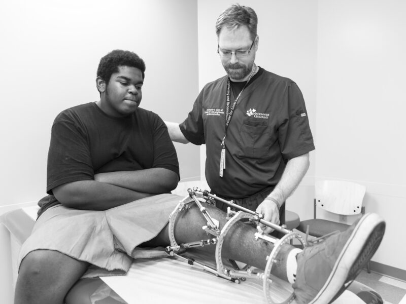
Distraction osteogenesis enables slow three-dimensional correction of deformity. Indications include a multiplane deformity with at least 5 mm of necessary length correction. This technique is challenging, and complications are many, including pin tract infections, skin tenting, dermatitis, need for repeat surgeries, and pain. It is important to evaluate for the psychological status of the patient and family and ensure appropriate candidacy for this undertaking, confirming a strong support network.
Ultimately, deciding the best treatment course when presented with a distal radius physeal bar is multifactorial. Age and remaining growth potential of the patient, the size and location of the bar, and patient and family expectations are key factors that should guide recommendations.
Reference:
Samora, J. Distal Radial Physeal Bar and Ulnar Overgrowth, Indications for Treatment, Epiphyseodesis vs. Ulnar Shortening Osteotomy in Adolescents. Pediatric Orthopaedic Society of North America Pre-Course. May 2020.
Image credit: Nationwide Children’s
About the author
Julie Balch Samora, MD, PhD, received her orthopaedic training at The Ohio State University and completed a fellowship in hand and upper extremity surgery at Harvard. She earned a Bachelor of Fine Arts at Carnegie Mellon University, a masters of music at Yale University, and an MD/PhD at West Virginia University, where she concomitantly earned masters degrees in public health as well as public administration.
Dr. Samora specializes in pediatric hand surgery at Nationwide Children’s Hospital, where she is Associate Medical Director of Quality, Director of Orthopaedic Quality Improvement, Director of Hand and Upper Extremity Research, and Director of the Clinical Quality Improvement Fellowship. Dr. Samora is on the board of directors of the Pediatric Orthopaedic Society of North America and is the current president of Ruth Jackson Orthopaedic Society and is committed to improving diversity in the field of orthopaedics. She is the Deputy Editor of AAOS Now and has published dozens of peer-reviewed manuscripts and text book chapters. Dr. Samora has a passion to provide safe, efficient, culturally-sensitive, and excellent patient care, focusing on best practices and quality improvement initiatives.
-
Julie Samora, MD, PhDhttps://pediatricsnationwide.org/author/julie-samora-md-phd/June 3, 2021
- Posted In:
- Features


