From Scan to Plan: Making Virtual Surgical Planning the Standard of Care for Ortho-oncology Operations
From Scan to Plan: Making Virtual Surgical Planning the Standard of Care for Ortho-oncology Operations https://pediatricsnationwide.org/wp-content/uploads/2024/09/080724BT85-crop-1024x281.jpg 1024 281 Abbie Miller Abbie Miller https://pediatricsnationwide.org/wp-content/uploads/2023/05/051023BT016-Abbie-Crop.jpg
“We deal in rarities,” says Thomas Scharschmidt, MD, director of the Pediatric Orthopedic Oncology Program at Nationwide Children’s Hospital and professor of Orthopedics at The Ohio State University. “When you consider that across the entire U.S. population, we have between 2,500 and 3,000 cases of primary malignant bone tumors each year, and only half of those are kids, every case requires unique consideration and planning.”
While in the past nearly every patient with a bone tumor would be facing amputation, modern limb salvage techniques combined with improving survival rates make orthopedic oncology an expanding field.
According to Dr. Scharschmidt, caring for children with sarcomas, the largest category of tumor seen by ortho-oncology experts, is a “team sport.” At Nationwide Children’s the sarcoma team includes representatives from orthopedic surgery, medical oncology, surgical oncology, plastic surgery, radiology, pathology, radiation oncology, physician assistants and nurse practitioners, as well as the legion of nurses and other clinicians supporting all of these areas.
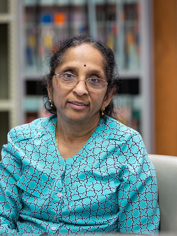
The team also includes Jayanthi Parthasarathy, BDS, MS, PhD, manager of the 3D Printing and Innovations Lab in the Department of Radiology at Nationwide Children’s. Dr. Parthasarathy is bringing the transformational impact of 3D printing and virtual modeling to patients across the hospital. Her collaboration with Dr. Scharschmidt has made it the standard of care for sarcoma patients.
Virtual Surgical Planning (VSP)
“The best analogy I can think of is this: We never get on a plane without a pilot who has a flight plan completely mapped out and in place before we get on the plane,” says Dr. Scharschmidt. “But I think we often do the opposite in surgery. We have a general idea of where we are going, but we do a lot of figuring out how to get there in the operating room. The idea behind VSP is to use the tools at our disposal to have that complete flight (surgical) plan in place for us, the families and the trainees, before going to the operating room. This makes the surgery more efficient — and safer for the patient.”
The first step to building the virtual model is to obtain magnetic resonance imaging (MRI) and computed tomography (CT) scans. The scans need to be taken on the same day with imaging markers placed to help guide image layering. The size of the image slices must also be 1 mm or smaller to ensure the accuracy of the resulting model.
“In the transition to making this part of our standard of care, we’ve moved the orders for these images into our electronic medical record,” says Dr. Parthasarathy. “This ensures every person, from the ordering physician to the technologists and radiologists, knows the requirements for a successful model.”
If the surgeons want to plan for a reconstruction using bone from another body part, the patient will also have a CT scan of that area.
Once she has the images, Dr. Parthasarathy builds the 3D virtual model and calls in the surgeons.
“We tend to see anatomy in 3D, and surgeons are very hands-on people,” says Dr. Scharschmidt. “Using the virtual model, we’re able to see that three-dimensional anatomy and plan our surgical margins more precisely.”
Once the surgeons have made their plan and marked their margins, Dr. Parthasarathy begins work on the physical models.
3D Printing: Reference Models
“Our multicolored, 3D printed models enable physicians to see a physical representation of their plan,” says Dr. Parthasarathy.
With sarcoma surgeries, surgeons are always removing part of the body — even in the case of limb salvage, she says. “This is hard for the patients. The models can help them see what will happen and why.”
“It’s really powerful for families and patients to see the planning,” agrees Dr. Scharschmidt. “It helps them better understand our goals of surgery before we get into the operating room.”
When it comes time for surgery, the 3D model heads to the operating room with the surgeon. While it stays outside the sterile fi eld, it’s there as a reference if needed.
“When figuring out resections and reconstructions in the operating room, there’s usually longer surgical time, with associated risks of higher anesthetic times and increased infection risk,” says Dr. Scharschmidt. “By utilizing VSP and 3D printing, we’ve shown we can do it better.”
In addition to the reference model, the VSP process enables the creation of custom implants and tools.
“We work with a vendor to use our models to create custom tools, such as cutting jigs to guide resections or custom implants,” says Dr. Parthasarathy. “The implants may include 3D printed metal components to replace portions of the pelvis or shoulder — complex areas where a patient-specific approach is essential.”
Proof Points
In 2022, Drs. Parthasarathy and Scharschmidt published a proof-of-concept description of their VSP and 3D printing process. The paper, published in the International Journal of Computer Assisted Radiology and Surgery, showed that the accuracy of the resections and the model predictions were within −0.29 to 0.45 mm (mean −0.09).
Since then, the team has continued to improve their process.
“We verify models by CT scanning the printed models,” Dr. Parthasarathy says. “In testing, we’ve shown a 0.01 to 0.05 mm range of variance — our models are incredibly accurate.”
The team has also continued to track the accuracy of the models for resection and reconstruction.
“To have the best possible impact on patient outcomes and surgical processes, we need to know if our models are accurate — not just anatomically accurate — but did we follow the plan we set, and if not, why?” says Dr. Scharschmidt. “This is an ongoing area of study, and we expect to publish our latest data toward the end of this year.”
This article appeared in the 2024 Fall/Winter issue. Download the issue here.
Reference:
Parthasarathy J, Jonard B, Rees M, Selvaraj B, Scharschmidt T. Virtual surgical planning and 3D printing in pediatric musculoskeletal oncological resections: a proof-of-concept description. International Journal of Computer Assisted Radiology and Surgery. 2023;18(1):95-104.
Image credit: Nationwide Children’s
About the author
Abbie (Roth) Miller, MWC, is a passionate communicator of science. As the manager, medical and science content, at Nationwide Children’s Hospital, she shares stories about innovative research and discovery with audiences ranging from parents to preeminent researchers and leaders. Before coming to Nationwide Children’s, Abbie used her communication skills to engage audiences with a wide variety of science topics. She is a Medical Writer Certified®, credentialed by the American Medical Writers Association.
- Abbie Millerhttps://pediatricsnationwide.org/author/abbie-miller/
- Abbie Millerhttps://pediatricsnationwide.org/author/abbie-miller/
- Abbie Millerhttps://pediatricsnationwide.org/author/abbie-miller/
- Abbie Millerhttps://pediatricsnationwide.org/author/abbie-miller/
- Posted In:
- Features
- Research
- Uncategorized


