A Key to Preventing Stenosis in Tissue-Engineered Vascular Grafts
A Key to Preventing Stenosis in Tissue-Engineered Vascular Grafts https://pediatricsnationwide.org/wp-content/uploads/2020/10/TEVG-Stenosis-.jpg 600 400 Kevin Mayhood https://secure.gravatar.com/avatar/bd57a8b155725b653da0c499ae1bf402?s=96&d=mm&r=g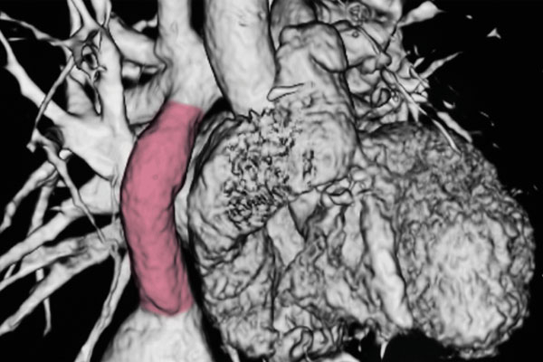
Seeding a high number of bone marrow mononuclear cells on graft appears to prevent narrowing.
Tissue-engineered vascular grafts hold promise for children with congenital heart disease because the grafts, which carry a patient’s own cells, have the potential to grow and regenerate just as the child’s blood vessels do. But over time, some grafts develop stenosis, or narrowing that can restrict blood flow.
Researchers at Nationwide Children’s Hospital suggest that the key to avoiding stenosis is seeding the graft with a large number of bone marrow mononuclear cells (BM-MNC). In a study using mouse models, 70 percent of grafts seeded with mononuclear cells isolated from peripheral blood developed stenosis compared to 11 percent of grafts seeded with cells isolated from bone marrow.
“One reason that bone marrow mononuclear cells may work is they seem to affect platelet function whereas other cells that we studied do not,” says Cameron Best, a PhD student and researcher in the Tissue Engineering Program at The Research Institute at Nationwide Children’s, and lead author of the study.
Best and colleagues found that BM-MNCs reduce blood platelet activation and accumulation that can result in graft thrombosis and progressive vessel occlusion. They found the higher the cell concentration, the more platelet activity was suppressed.
They report their findings in the journal ACS Biomaterials Science & Engineering.
Congenital heart defects are the most common birth defect and despite advances, are the leading cause of death among newborns.
“One source of problems is the manmade materials needed for vascular grafts or replacement heart valves to correct congenital heart defects,” says Christopher Breuer, MD, director of the Center for Regenerative Medicine and senior author of the study. Grafts using Gore-Tex or Dacron are prone to infection, thrombosis and stenosis. Because they don’t grow, the child may need to undergo a series of operations to replace outgrown grafts.
Dr. Breuer has been working on tissue-engineered grafts for two decades. This study looked at the seeding process his lab has developed, and which is currently being used in the first phase of an FDA-approved clinical trial, to determine which steps may reduce the risk of stenosis.
The research team found that application of a phosphate-buffered saline solution they use to pre-treat the biodegradable scaffold and the step of incubating the seeded scaffold in plasma from the mouse failed to reduce stenosis. Only the presence of BM-MNCs appeared to keep the grafts open — in nearly 90 percent of cases.
The researchers then tested whether the more easily accessible mononuclear cells circulating in the bloodstream would perform as well. They didn’t.
While Dr. Breuer and Best continue to study the mechanisms by which the BM-MNCs may reduce chances of stenosis, the researchers will also test second-generation grafts that will carry more of the cells. “We’ve completed a lot of work on harvesting more bone marrow mononuclear cells and getting more cells seeded on the scaffold,” Dr. Breuer says.
The study has also helped the researchers streamline their process. After showing that incubation in plasma adds nothing to the outcomes, that time-consuming step has been removed.
The grafts are made while a patient is in the operating room and he or she will spend less time under anesthesia.
Dr. Breuer says the goal of the work hasn’t changed for 20 years. “Ultimately, if we can develop methods to rapidly produce blood vessels, valvular patches and heart valves without stenosis, we can have a positive impact on children with congenital heart disease.”
Reference:
Best C, Tara S, Wiet M, Reinhardt J, Pepper V, Ball M, Yi T, Shinoka T, Breuer C. Deconstructing the Tissue Engineered Vascular Graft: Evaluating Scaffold Pre-Wetting, Conditioned Media Incubation, and Determining the Optimal Mononuclear Cell Source. ACS Biomaterials Science & Engineering.2017;3(9):1972-1979.
Photo credit: Nationwide Children’s
About the author
-
Kevin Mayhoodhttps://pediatricsnationwide.org/author/kevin-mayhood/

-
Kevin Mayhoodhttps://pediatricsnationwide.org/author/kevin-mayhood/
-
Kevin Mayhoodhttps://pediatricsnationwide.org/author/kevin-mayhood/
-
Kevin Mayhoodhttps://pediatricsnationwide.org/author/kevin-mayhood/